Ceramide
2022-08-11: reference:
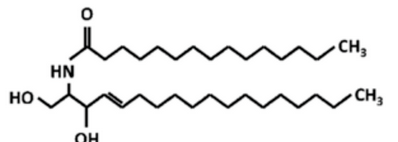
Ceramide
 #
#
-
-
-
 is this comprehensive, or what? Not sure
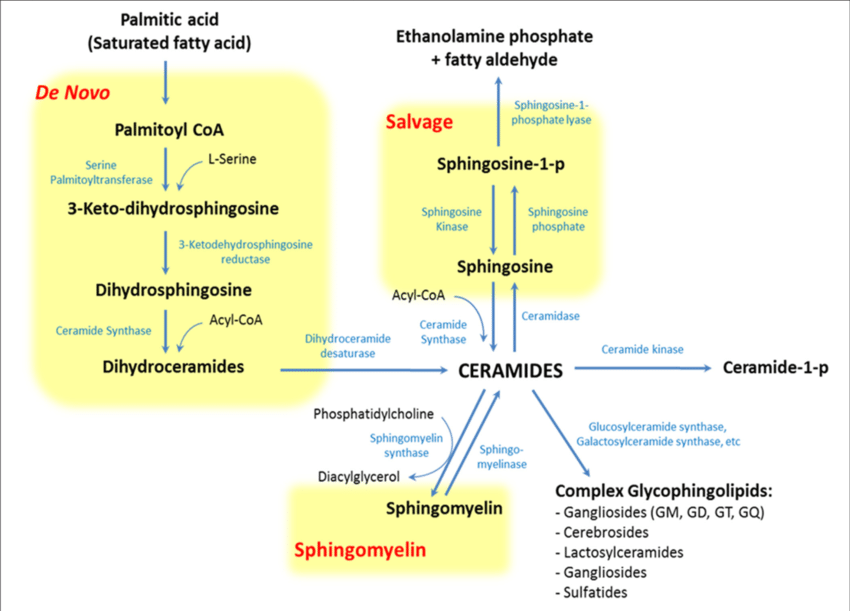
 - (They skipped 3-keto-dihydrosphingosine -> Sphinganine)
- There are 6 Ceramide Synthase subtypes each with fatty acid specificity:
- CerS1: Specifically Stearic Acid. Basically only in brain and skeletal muscle.
- CerS2: C20-26. Highest expression of all CerS in oligodendrocytes/Schwann cells.
- Indeed, this de novo synthesis of ceramide is the rate-limiting step of the synthesis of all the complex sphingolipids, sphingomyelin, and so on.
-
-

-
In schizophrenia’s structural sphingolipid abnormality, myelin-associated glycoprotein and myelin/oligodendrocyte glycoprotein are segregated from the lipid raft.
-
-
Similarly, The Role of Ceramides in Insulin Resistance (2019)
-
Ceramide function in the brain: when a slight tilt is enough
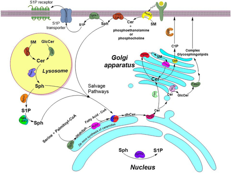
- De novo synthesis of ceramides takes place on the cytoplasmic face of the Smooth Endoplasmic Reticulum and in mitochondria.
-
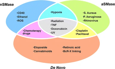 all these things stimulate. The study describes most of these things as ‘inducers of cell death’. Ceramide production results in cell deaht.
-
Ceramide channels and mitochondrial outer membrane permeability
- They permeabilize the Outer Mitochondrial Membrane
- The dogma in early mitochondrial research was that channels had to be small and highly selective.
- It is uncertain in which direction the influence is, but it synergizes with BAX in permeabilizing the MOM.
-
Bioactive sphingolipids in the modulation of the inflammatory response
- SphK1 and CERK1 are probably effectors of cytokine-mediated Prostaglandin generation in inflammatory conditions.
- It was shown that S1P pathway but not C1P pathway mediated IL-1β-mediated COX-2 induction.
- Ceramide-1-phosphate activates cPLA2.
- PLA2/COX-2 colocalized with the golgi and perinuclear regions in response to S1P/C1P treatment.
- LPS treatment produces rapid rise of ceramide in Macrophages. LPS as well as ceramide analogues induce TNF-α secretion. However, the fact that LPS but not ceramide analogues stimulated interferon activity suggested that ceramide induced only a subset of LPS-induced genes.
- SphK1 and CERK1 are probably effectors of cytokine-mediated Prostaglandin generation in inflammatory conditions.
-
Ceramide-1-phosphate (via ceramide kinase). This facilitates localization of p-Phospholipase A2 to the trans-Golgi
- Indeed it locazlies to the COX-2-localization site on the Outer Mitochondrial Membrane
-
Autophagy in the light of sphingolipid metabolism
- Promotes Ca2+-dependent Liposomeal fusion, enhancing vesicle fusion.
- C1P levels increase during phagocytosis, indicating that C1P may promote fusion between phagosomes and lysosomes
Neurodegenerative Disease #
-
Triangulated mal-signaling in Alzheimer’s disease: roles of neurotoxic ceramides, ER stress, and insulin resistance reviewed.
- Ceramide accumulation appears to be bidirectional with oxidative/ER stress and neuroinflammation.
-
Amyloid-β peptide induces oligodendrocyte death by activating the neutral sphingomyelinase–ceramide pathway
- Activates neutral sphingomyelinase - but not Acid Sphingomyelinase…
- Blocking ceramide degradation with Oleoylethanolamine exacerbated Aβ cytotoxicity.
- Addition of sphingomyelinase (from bacteria) induced Oligodendrocyte death.
- Astrocytic ceramide as possible indicator of neuroinflammation (In frontotemporal lobar dmentia Pick’s disease)
- Association between Plasma Ceramides and Phosphatidylcholines and Hippocampal Brain Volume in Late Onset Alzheimer’s Disease
- I swear they can be as short as 14 carbons or as long as 26.
- Aging-dependent mitochondrial dysfunction mediated by ceramide signaling inhibits antitumor T cell response
Phytoceramide #
- Phytoceramide has an extra hydroxyl group.
- Administration of Phytoceramide Enhances Memory and Upregulates the Expression of pCREB and BDNF in Hippocampus of Mice